Review Article | | Peer-Reviewed

Magnetic Resonance Imaging in the Detection of Neuroanatomical Changes in Down Syndrome: A Narrative Review from the Developing Fetus to Childhood

Received: 31 March 2025     Accepted: 9 April 2025     Published: 19 May 2025
Views:       Downloads:
Abstract

Background: Down syndrome (DS) or trisomy 21 is a genetic disorder caused by unusual cell division and an extra copy of chromosome 21. DS is characterized by phenotypic characteristics identified clinically and is the most common genetic cause of intellectual disability in children. DS can also result in other medical complications that involve the heart, the digestive system and memory disabilities leading to Alzheimer’s disease (AD). Although diagnosis is made clinically, screening tests such as chorionic villus sampling (CVS), amniocentesis and ultrasound detecting translucency of the nuchal folds of the neck are used to detect it at its early stages. There is no known treatment for DS, however, early detection and intervention can improve quality of life (QoL) for patients and their parents. Aims: This review aims to identify the importance of neuroimaging, particularly magnetic resonance imaging (MRI), in early diagnosis of DS. The review will explore how neuroanatomical changes can guide future research and focus interventions to target needs of individuals. Method: This review included 12 studies on DS from the year 2000 to date. Participants included ranged from fetus to 15-year-old teenagers. Diagnosis of DS was first made by CVS, amniocentesis or ultrasound depending on mother’s preference followed by karyotyping confirming trisomy 21. The main imaging modality included in the review is high-resolution MRI which all participants underwent for comparison. Results: Results of the MRI showed an overall reduction in volume in different areas of the brain in DS patients compared to controls. Most prominent volume reduction were found in the frontal lobes, hippocampus and brainstem. With increasing age there was preservation of volumes in parietal and temporal lobes. After the age of 11 years, changes in the grey and white matters started to appear. Conclusion: Early detection of brain changes, especially in fetus, could improve developmental outcomes for people suffering from DS by providing early and tailored interventions focusing on potential cognitive impairments associated to damaged brain areas. These neuroanatomical changes in DS patients are correlated to cognitive disabilities that are controlled by specific areas of the brain, relating this review to its clinical relevance. Some limitations of the studies included in the review was the small sample size. Selection bias was introduced by recruiting participants solely from hospitals where healthcare access might be limited to those can afford it. Some strengths include the consistency in diagnosing DS first with chorionic villus sampling, amniocentesis and ultrasound and confirmation it with karyotyping.

Published in American Journal of Pediatrics (Volume 11, Issue 2)
DOI 10.11648/j.ajp.20251102.18
Page(s) 81-92
Creative Commons

This is an Open Access article, distributed under the terms of the Creative Commons Attribution 4.0 International License (http://creativecommons.org/licenses/by/4.0/), which permits unrestricted use, distribution and reproduction in any medium or format, provided the original work is properly cited.

Copyright

Copyright © The Author(s), 2025. Published by Science Publishing Group

Keywords

Down Syndrome, Trisomy 21, Developing Fetus, Childhood, Magnetic Resonance Imaging, MRI, Cognitive Disabilities

1. Introduction
Down syndrome (DS) also known as trisomy 21 is a genetic disorder caused by inheriting extra allele of chromosome 21 characterized by phenotypic manifestations (see Figure 1) . Environmental risk factors for developing DS include tobacco use, folic acid supplementations and oral contraceptives . The most important risk factor for DS is advanced maternal age, which results in non-disjunction of homologous chromosomes during cell division known as meiosis . DS is diagnosed during antenatal period via ultrasound in the first trimester looking for fetal nuchal translucency, which is the pouch of fluid present behind the neck of the fetus and during the second trimester by looking at alpha-fetoprotein, B-HCG, estriol and inhibin-A in maternal serum and amniotic fluid . To date no treatment for DS have been found, however genetic counselling and behavioural interventions have been approved to increase the quality of life (QoL) for parents and their child .
DS is one of many neurodevelopmental disorders that can cause changes to developing brain early in fetal life caused by extra allele of chromosome 21 . Therefore, neuroimaging techniques brought the possibility to advance our knowledge in the field of neurodevelopmental disorders, shedding light on neurological changes behind the distinctive phenotype observed in DS. For instance, high resolution magnetic resonance imaging (MRI) is the most used neuroimaging technique in the study of DS for its ability to detect volume changes in specific areas of the brain . MRI provide basis for pathogenesis of DS by increasing understanding of cognitive challenges that can be faced by DS patients depending on what neuroanatomical changes are observed on their image (see Figure 2). Neuroimaging provides valuable insights into brain changes in DS, enabling personalized interventions for cognitive, motor, and speech development. It also helps healthcare providers and caregivers improve communication, manage behaviours like aggression and early detection of Alzheimer’s disease (AD). Additionally, neuroimaging advocates for resources, reduces stigma and enhance the overall QoL for individuals with DS and their families . This review aims to explore and summarise main neuroanatomical changes detected on MRI in patients with DS from developing fetus to childhood. This review highlights importance of early disease detection and intervention in improving QoL and cognitive abilities in DS. It also serves as a guide for future research to focus on other anatomical changes using other imaging modalities. The review provides better understanding of correlation between anatomical changes detected and cognitive abilities, thus healthcare providers can use this information to help them implement a more effective, individualized intervention that support the child's development and improve behaviour. Finally, caregivers can use information provided to better understand their child’s unique cognitive and emotional processing, allowing them to tailor strategies which will promote positive behaviour and enhance communication.
Figure 1. Phenotypic characteristics in Down syndrome .
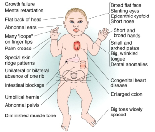
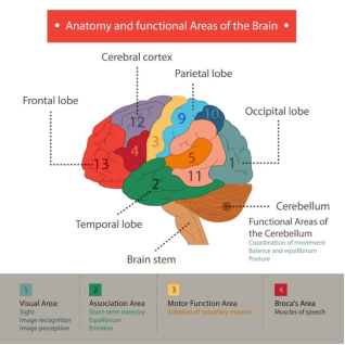
Figure 2. Areas of the brain and their related cognitive function .
2. Method
2.1. Search Strategy
To identify relevant literature on brain anomalies and changes in DS from fetal to childhood, a systemic review was conducted using several databases including PubMed, Scopus, and Google Scholar. Search strategy was structured around specific key words such as “Down syndrome brain anomalies”, “Neurodevelopmental changes Down syndrome”, “Fetal brain development Down syndrome”, “MRI brain changes Down syndrome”, Cognitive development Down syndrome childhood”, “Neuroanatomical changes in Down syndrome from fetus to childhood” and “Down syndrome brain changes from fetus to childhood”. A combination of keywords such as “Neuroanatomy AND Down Syndrome” and “Neurodevelopment AND Down Syndrome” was also used.
2.2. Inclusion Criteria
Results were filtered for articles published in the last 24 years to ensure only up to date articles included. Some articles were also acquired from references lists in review papers. Inclusion criteria included male and female children diagnosed with DS from fetus to 15 years old. Research found that after age of 15, changes in the brain tend to plateau and age-related changes start to appear . For example, DS patients after the age of 15 are at increased risk for developing AD . Only human based research and peer-reviewed original articles were included. The main outcome was MRI studies that detected neuroanatomical differences relevant to patients with DS.
2.3. Exclusion Criteria
Review papers, meta-analysis, opinion-based articles, editorials and non-scientific articles were excluded. Studies that used neuroimaging modality other than MRI were excluded. Papers that focused on metabolic changes, alterations in neurotransmitters and not on neuroanatomical changes were also excluded. Patients diagnosed with DS on any type of neurological treatment or suffering from other neurodevelopmental disorders were excluded.
2.4. Screening
Screening process started with titles that seemed relevant to the topic which included words like Down syndrome and neuroanatomical changes. Abstract reading was part of the initial screening process which helped exclude papers that did not use imaging modality, or which was outside the age range desired for the review. After the initial screening, full-text screening process included reading full articles and exclude papers irrelevant to the topic, or which did not meet the inclusion criteria.
2.5. Study Selection/Characteristics
Screening process started with 176 articles based on titles and abstracts. Any paper with no full text, no imaging modality, was not a neuroanatomical study or was a duplicate was removed. After reviewing titles, abstracts and removing duplicates, 150 articles remained for full-text screening. Full text screening excluded reviews, papers published before 24 years, ages above 15 years, any animal-based research, no imaging and papers with therapeutic outcomes. From 150 articles, only eight were included. Second, all 42 reviews were screened again and a total of 4 original articles were retrieved from references which adds up to the studies included in the review to a total of 12. The screening process was conducted by a single researcher and is outlined in figure 3.
2.6. Data Synthesis
This review used Popay et al., 2006 handbook as a guidance for data synthesis. The main elements of data synthesis as per Popay et al. are:
1. Developing a theoretical model of how interventions work, why and for whom
2. Developing a preliminary synthesis
3. Exploring relationships between data
4. And, assessing robustness of synthesis product .
3. Ethics
One of the main challenges was getting informed consent for minors from guardians. Before obtaining guardian’s consent, all information should be provided in detail and explained when in doubt. All studies in this review obtained a written consent from guardians that included all information on disease course, how MRI is conducted and any potential risks.
In addition, research that does not address gaps in knowledge and would not benefit the child and their family should be rejected to avoid unnecessary intervention on a vulnerable population . In all studies included in this review, imaging was carried based on current guidelines and regulations ensuring not only welfare of participants but also consistency. In the Fujii et al. study, images were taken by a paediatric neuroradiologist with 13 years of experience . As per guidelines, children and young patients should have high quality hospital care delivered by staff who have the right set of skills or under supervision by a higher qualified physician . According to MRI protocol for taking a brain image, axial T-2 weighted, coronal T1-weighted or sagittal T1 weighted images should be used in paediatric patients which was used by all studies in the review. Carducci et al. for example used an axial T1-weighted image, while Śmigielska-Kuzia et al. used coronal and sagittal T1-weighted imaging. It is also important that the review does not reinforce negative stereotyping or contribute to increasing stigma in the community towards individuals with DS. Paying attention to cultural sensitivity and psychological distress on families should be considered carefully. For example, In Tauri et al. study, all pregnant women who were reluctant to having amniocentesis or CVS were offered karyotyping after birth of the baby as an alternative to minimize distress that will result from undergoing an unwanted procedure.
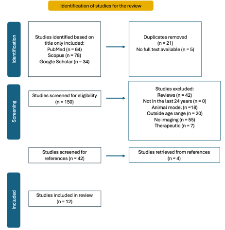
Figure 3. PRISMA flowchart showing screening method adapated for the review .
4. Results
4.1. Overview
All studies in the review shared the aim of exploring neuroanatomical changes that occur in brains of DS patients. The main aim was to establish relationship between anatomical changes and cognitive abilities of individuals with DS.
In 10 studies, a diagnosis of DS was made by prenatal ultrasound, CVS or amniocentesis followed by a confirmatory karyotyping which revealed extra allele of chromosome 21, or trisomy 21. However, in Yun et al. and Tarui et al. studies, researchers diagnosed DS patients by cell-free DNA followed by confirmatory karyotyping . Cell-free DNA testing is a non-invasive procedure which takes mother’s blood containing DNA with mother components and 10-20% fetal components. If the fetus has DS, there will be a slight increase in number of chromosome 21 specific DNA in maternal blood. This procedure can be performed in early pregnancy to reduce risk of non-invasive testing with CVS or amniocentesis . All participants were also tested for other neurological or neurodevelopmental diseases. Three of the studies in the review took measurements of intelligence quotient (IQ) scale and excluded participants with IQ levels less than 70. Hamadelseed et al. and Menghini et al. used the Stanford Binet Intelligence scale 5th edition (TBSS) . The other study that measured IQ was Carducci et al. using the Revised Wechsler Intelligence Scale for Children (WISC-R) . Measuring IQ levels is important because children with DS have varying degrees of intellectual disability and IQ levels can help quantify level of cognitive impairment and provide baseline that researchers can use for method standardization .
Neuroanatomical changes were studied using MRI and comparison of results between DS patients and healthy controls was obtained. ten of twelve studies used structural MRI solely for neuroimaging. The other two studies included the Fujii et al. study who used a diffusion weighted imaging (DWI) in addition to structural MRI and Gunbey et al. who used only DWI. DWI is useful in detecting changes in myelination process of white matter in the paediatric population through changes in water diffusion . The power of magnetic field of scanners measured in Teslas (T) and MRI sequence also differed between studies (see Table 1).
Gunbey et al. mentioned use of sedation before putting their 2-year-old patients into the MRI machine. They used 50 mg/kg of Chloral hydrate (CH) as per child’s regulations of using sedatives . Carducci et al. also used sedation for their 10-year-old patients but no mention of what type of sedation used was included . Pharmacodynamic of Chloral hydrate differs between children which introduces bias related to physiological differences in how different group of people respond to the drug. For example, the half-life of the drug is 9.7 ± 1.7 hours in toddlers but 39.8 ± 14.3 hours in infants . There have also been ethical issues raised regarding the drug because of its low therapeutic index, which is the dosage range between a medication’s effective and toxic dosages . The drug has been found to cause potential hepatic injury, cardiotoxicity and even death in some patients .
Summary of the results is demonstrated in Table 1.
Table 1. Summary table of papers included in the study including important information on methodology and results.

Study

Age

Total number of participants

MRI sequence (T1 or T2-weighted image)

Power of the magnetic field measured in Tesla (T)

Results

(Patkee et al., 2020)

21-46 weeks gestation

124

T2- weighted image

1.5T

3.0T

Reduced cerebellar volume in 2nd trimester

Alteration in cortical growth in 3rd trimester

(Kitano et al., 2023)

22-37 weeks gestation

22

T2- weighted image

1.5T

Small cerebellar vermis

Large fourth ventricle

Small cerebellar:fourth ventricle ratio

(Tarui et al., 2019)

25-29 weeks gestation

21

T2- weighted image

1.5T

Decreased cortical volume growth

Decreased subcortical parenchymal volume

Decreased cerebellar hemispheric volumes

(Yun et al., 2020)

29 weeks gestation

26

T2- weighted image

1.5T

Decrease sulcal depth in bilateral Sylvian fissures, right central and parieto-occipital sulci

Increased sulcal depth in left superior temporal sulcus

(Fujii et al., 2017)

0-11 years

64

T1- weighted image

T2- weighted image

1.5T

3.0T

Smaller ventral pons

(Gunbey et al., 2016)

2 years

18

T1- weighted image

1.5T

Reduced grey matter volumes of left putamen, thalamus, caudate nucleus, cerebellar cortex, brainstem and corpus callosum

Reduced subcortical grey matter and total cortical grey matter of both hemispheres

Decreased volume of right cerebellar white matter

(Shiohama et al., 2019)

< 3 years

40

T1- weighted image

3.0T

Decreased brain volumes in the grey and white matter, brainstem and cortical areas of the brain

(Śmigielska-Kuzia et al., 2011)

6 years

8 years

49

T1- weighted image

T2- weighted image

1.5T

Reduction in total brain volumes, frontal lobe and temporal lobe volumes

Reudction in volumes of the hippocampus and amygdala

(Carducci et al., 2013)

10 years

48

T1- weighted image

1.5T

Reduction in total brain volume

Reduction in grey matter of frontal lobes, white matter of frontal and parietal lobes and brainstem

Preservation of grey matter of parietal and temporal lobes and white matter of temporal lobe

(Pinter et al., 2001)

11 years

31

T1- weighted image

1.5T

Smaller overall brain volume

Small cerebellar volume that is disproportional to larger subcortical grey matter volume

Preservation of parietal lobe grey matter

Preservation of temporal lobe white matter

(Hamadelseed & Skutella, 2023)

14-15 years

25

T1- weighted image

1.5T

3.0T

Mean total brain volume 20% smaller than the control group

No difference in parietal lobe, dentate gyrus, fusiform gyrus and parahippocampal gyrus

(Menghini et al., 2011)

15 years

24

T1- weighted image

1.5T

Reduced total brain volumes (TBV), grey matter volume (GMV) and white matter (WMV) volume

4.2. MRI Changes in the Developing Fetus
Patkee et al. included fetuses with mean age 28.6 weeks diagnosed with DS and compared them to controls of mean age 30.6 weeks. Comparison was also done on 36-46 weeks neonates with DS and 38-46 weeks controls. There was a reduction in cerebellar volume that started in the second trimester for DS fetuses and neonates. In comparison, fetuses and neonates in the control group started showing reduction in the third trimester . DS patients also exhibited a Cerebellar volume that is disproportionally smaller than whole brain volumes . Similarly in another study, cerebellar to fourth ventricle ratio was smaller in DS patients . Researchers also observed a reduction in vermis height and surface area in the second trimester and a reduction in transcerebellar diameter and vermis width in the third trimester . Kitano et al. similarly found a reduction in the height (HV) and anteroposterior diameter (APDV) of the cerebellar vermis in DS fetuses .
Cortical growth changes in fetuses and neonates with DS revealed a smaller occipitofrontal diameter (OFD), which is the distance between occipital and frontal lobes, in second and third trimesters. A smaller skull occipitofrontal diameter (skull OFD), which is the distance between frontal and occipital skull bones was also evident and a smaller head circumference (HC) was detected in in the third trimester compared to controls . Another study detected similar decreased growth of cortical plate, subcortical parenchyma and cerebellar hemispheres beginning at 28 weeks. There was also less growth in their inner cerebral surface area of the right hemisphere compared to controls . Their findings are consistent with previous research on autopsy which detected decreased cerebral and cerebellar sizes in the brains of DS fetuses .
Larger lateral ventricular volumes was also detected in DS patients by two studies . On the other hand, Tarui et al. detected no ventriculomegaly on imaging, defined by atrial width more than 10-mm in coronal plate . This difference could be a result of a smaller sample size in Tarui et al. study with 21 participants compared to 124 participants in the Patkee et al. study. Also, while the range of ages in the Tarui et al. study was limited at 29 weeks, both Patkee et al. and Kitano et al. studies included participants up to 46 and 37 weeks, respectively. Therefore, ventriculomegaly changes could have not been detected by Tarui et al. because of small sample and because these changes are only detectable in older weeks.
Another study underwent a different approach that focuses on cortical folding early in fetal life. They argued that despite a lot of research being conducted on different brain volumes, little research is done on how abnormal cortical folding early in development can lead to impairment in cognitive function, brain malformations and neurodevelopmental disorders. Therefore, studying abnormalities in cortical folding during the fetal development will add more knowledge to our understanding of developmental disorders. Researchers included fetuses with DS at 29 gestational weeks and compared them healthy age-matched controls. On average patients with DS had lower whole brain sulcal depths more pronounced in the bilateral Sylvian fissures, right central and parieto-occipital sulci. However, increased sulcal depth in the left superior temporal sulcus was the cause of the atypical cortical folding and hemispheric asymmetry. Those differences in sulcal depth increased with gestational age . Therefore, sulcal depth is considered an important marker for studying early brain development.
4.3. MRI Changes from 2 to 11 Years Old
In a study, 2 years old children with DS showed reduction in grey matter volumes of left putamen, thalamus, caudate nucleus, cerebellar cortex, brainstem and corpus callosum. There was also reduction in subcortical grey matter and total cortical grey matter of both hemispheres. In addition, lower volume of right cerebellar white matter was also observed . While no significant difference was found in total white matter and grey matter in the Gunbey et al. study, another study showed decreased brain volumes in DS patients in the grey matter bilaterally, right white matter, brainstem, right superior temporal cortex (STC), right rostral anterior cingulate cortex (ACC) and left rostral middle frontal cortex (MFC) . Potential reasons for the difference include a smaller sample size, the use of sedatives which can introduce bias into the results and the additional use of DWI by Gunbey et al. compared to Shiohama et al study. Research supports that structural MRI is better at detecting anatomical details and provides a stronger grey/white matter contrast compared to other scans . Research also concluded that higher magnetic power is better at detecting changes and while Shiohama et al. used a 3.0 T scanner, Gunbey et al. used a 1.5 T scanner .
Volumes of the hippocampus, amygdala, temporal and frontal lobes were investigated in 6 year old children with DS and 8 years old controls. Results showed a reduction in total brain volume in DS patients by 13% . Supported by a study of 10-year-old patients which showed reduction in total brain volume as well as the grey matter of frontal lobes, white matter of frontal and parietal lobes and the brainstem . A reduced volume in right frontal lobe by 24%, left frontal lobe by 25.2%, amygdala by 23.5%, right temporal lobe by 18.6% and left temporal lobe by 14.2% was also detected . Another study showed no significant difference in frontal lobe, occipital lobe and superior temporal gyrus volumes compared to controls . Research has shown that the use of T1-weighted imaging combined with T2- weighted imaging is superior to using any of the two sequences alone for better mapping and discrimination between brain regions and detection of structrual impairments . Between the two studies, the combination of T1 and T2 sequences used by the Śmigielska-Kuzia et al. study allowed for better detection of changes in the frontal lobes compared to the use of T1 sequencing alone in Pinter et al. study.
Almost all studies found preservation of parietal lobe volumes in DS patients. Pinter et al. argued that even though parietal lobe white matter volumes and temporal lobe white matter volumes were larger compared to controls, no significant difference was found between parietal lobe grey matter, temporal lobe grey matter and superior temporal gyrus volumes. Preservation of grey matter in the parietal lobe is consistent with the strength in visuospatial skills observed in DS patients . Other researchers also reported preservation of grey matter of parietal and temporal lobes volumes and white matter of temporal lobe volume in DS patients . However, the most significant volume reduction was in the hippocampus by 38% . This is because the hippocampus which is located deep in the temporal lobe is responsible for memory and learning both of which are severely affected in patients with DS .
4.4. MRI Changes in Childhood from 11 to 15 Years of Age
Similar to previous results of 6 years and 10 years old patients, a study of 15 years old DS patients had total brain volume that is 18% smaller with a mean volume of 1068.3 cm3 compared to 1297.5 cm3 . In another study of 15 years old patients with DS showed reduced total brain volumes (TBV), grey matter volumes (GMV) and white matter volumes (WMV), see table 2 .
Table 2. Total volume of brain regions in 15-year old patients with DS compared to controls .

Region

Calculated volumes in cubic cm

Down syndrome

Controls

Grey matter (GM)

677.3 cm3

783.6 cm3

White matter (WM)

373.8 cm3

441.2 cm3

Total brain volume (TBV)

1443.6 cm3

1628.7 cm3

While most changes before this age were in the cortical regions and cerebellar lobes, imaging in this age group started detecting changes in the whole GM, WM and TBV that were not present before this age. As demonstrated in Table 2 there is a total difference by 106.3 cm3, 67.4 cm3 and 185.1 cm3 in the GM, WM and TBV respectively in DS patients . Figure 4 also shows the difference in volume density in specific areas of the brain.
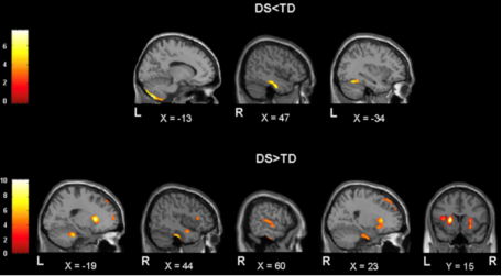
Figure 4. Difference in volume densities in 11 to 15 years of age patients with DS compared to controls in different parts of the brain. The top part of the figure shows controls with more grey matter volume illustrated by increased densities in some areas of the brain compared to increased grey matter densities detected in DS patients in other brain regions shown at the bottom. Overall, there is a reduction in volumes of left posterior cerebellum, right inferior temporal gyrus, right and left medial temporal lobes in DS patients .
In DS Patients certain areas of the brain detected larger grey matter volume including the left anterior cerebellum, right medial temporal lobe, right and left basal ganglia, right and left insula, left and right superior frontal gyrus, right superior and middle temporal gyrus and left and right inferior frontal gyrus all shown in figure 4 . By contrast, In another study 15 year old DS patients showed no significant difference in the volumes of the parietal lobe, dentate gyrus, fusiform gyrus and para-hippocampal gyrus. However, MRI results of the whole brain revealed a total brain volume that is 20% smaller in DS patients . Both researchers then compared their results to how they correlate to cognitive abilities of DS patients.
Menghini et al. detected that reduced volume densities correspond to morphosyntactic comprehension and production (cerebellum and left temporal gyrus), lexial comprehension (cerebellum) and visuo-perceptual abilities (frontal gyrus). Therefore, patients with DS are expected to have difficulties in those cognitive abilities. While results of short- and long-term memory correspond to areas of the brain that exhibited increased volumes such as the parietal and right temporal gyrus. Therefore, only some patients with DS may exhibit dysfunction in memory progressing to AD . Similarly, Hamadelseed et al. results revealed similar correlation between difference in brain volume and cognitive abilities in DS patients . Both results confirm what previous researchers found between brain volumes and cognitive functions in DS patients .
5. Discussion
5.1. Summary of Synthesis Results
This review provides insight into the developmental differences between individuals with DS and healthy controls from the fetus to childhood. A key finding across all studies is the reduction in brain volume in the cerebellum, cortical and subcortical regions. Reduction in the size of the cerebellar vermis and delays in cerebellar maturation was also observed in some studies. These changes are consistent with other research which observed reduced cerebellar size in DS patients during autopsy .
In addition, cortical growth delay was observed in the second trimester , consistent with reduction in cortical grey matter and subcortical structures observed at later stages . These changes are also consistent with postmortem autopsy findings, emphasizing that those are not imaging artifacts, but they reflect true neurodevelopmental changes .
Most importantly, preservation of parietal lobe grey matter volumes was observed by Pinter et al. and Carducci et al. studies. This was accompanied with preservation of visuospatial abilities in DS patients as the parietal lobe plays a key role in this cognitive function. Therefore, preservation of the parietal lobe and possibly other areas is an important field of research that needs to be investigated further. Findings by Carducci et al. and Pinter et al. also found preservation of temporal lobe grey matter but a reduction in frontal lobe volumes. These differences reflect patterns of cognitive strengths and weaknesses in DS where certain functions such as visuospatial processing controlled by the temporal lobes may be less affected than others such as memory and language controlled by the frontal lobes.
Abnormalities in sulcal depth early in development also highlights the importance of abnormal cortical folding observed in DS . However, more research is needed on the topic as it highlights important differences in the neurodevelopmental stages early in life which could be used as a guidance for an early targeted intervention.
With increasing age, research found that changes in neurodevelopmental started to appear in places such as the hippocampus, amygdala and frontal lobes . The hippocampus showed the most significant reduction out of all brain volumes affected, which is an important finding due to its role in memory and learning .
Changes in regions of GM, WM and TBV was most prominent in childhood. GM changes reflect changes in learning and memory observed in DS patients, while changes in WM which contains myelinated axons that allows for communication between different brain regions is reflected by changes in motor and cognitive abilities. While changes in frontal lobe and hippocampus are reflected by changes in executive functions like planning, decision making, problem solving and memory.
The transition from early cerebellar and cortical alterations observed before childhood to more global brain atrophy starting at the age of 11 onwards is crucial for understanding long-term progression of the disease and cumulative impact of neurodevelopmental changes on cognitive functions. Given the progressive nature of neurodevelopmental diseases, future research should focus on early interventions that act on neuroplasticity to reduce cognitive impact.
5.2. Strengths and Limitations of Studies
One important limitation across all studies is the small sample size which decreases statistical power and therefore cannot be attributed to the whole population. In addition, money and time were allocated to the research and for it to be done on a small sample that will not contribute to improvements in clinical practice might be considered as wrong allocation of resources . Also, selection bias was introduced by all studies because all participants were recruited from hospitals. The problem with this selection is that it does not reflect the whole population but only those who are able to access health care facilities because of greater wealth or are living in closer proximity to the center . The different use of magnetic field power between studies could also lead to bias in group comparisons. Images acquired at a stronger magnetic field benefit from the increased power by obtaining better resolution and reconstruction of images. In the Kitano et al. study, some participants suffered from congestive heart failure (CHD) and because of limited resources were unable to assess the impact that CHD could also have on fetal brain growth unrelated to DS. However, an important strength is the use of living human brains of DS patients to be imaged and analysed across all studies. An important strength that allows for this review to be implemented in the population.
5.3. Limitations of Review
This review used single imaging modality in investigating neuroanatomical changes. In studying brain disorders the use of one imaging modality has limited interpretability and applicability, while multi-modal imaging enhances one’s understanding of the neuropathological processes that occur in patients with DS and provides improvement in clinical data and research . There was also no consideration for co-morbidities that may exist in patients with DS in this review. Patients with DS are susceptible to congenital heart diseases, autism, depression and dementia. These co-morbidities may cause neuroanatomical changes that are not related to DS .
5.4. Guidance for Future Research
Combination of structural, function and molecular imaging techniques for better understanding of pathology in DS. To date, most literature used one single imaging modality in studying the pathological processes that occur in the brains of DS patients. However, as DS is a multifactorial disease with multiple pathological processes occurring at the same time, future research should integrate the use of combination of neuroimaging techniques to understand the pathological process that interplay in the developmental process in those patients. Combination of neuroimaging data allows for consideration of different biological mechanisms, for example, brain metabolism can be detected from positron emission tomography (PET) scans, neurotransmitters and metabolite levels are examined by spectroscopy and structural imaging can be used to detect volumetric changes. It is also important to consider special biomarkers and their role in advanced DS specifically amyloid proteins and neurofibrillary tangles features that are found in progressive DS that results in Alzheimer’s disease . Neuroimaging models can also be used to address gaps in research in areas such as gene silencing, which could be a promising in the field of neurodevelopmental disorders.
5.5. Implementation in Society
This review provides valuable insights into neuroanatomical changes in DS that can help improve QoL for both patients and their caregivers. MRI can reveal specific areas of the brain that are affected to help create a personalized plan for cognitive, motor or speech development. Understanding the root cause of behavioural changes can guide caregivers refine strategies that will enhance communication with their child and reduce reliance on nonverbal cues. It also provides information for improving emotional well-being of patients to reduce aggression and anxiety not only towards their parents but also with their siblings, peers and adults. Identifying areas where the brain is affected can optimize cognitive training and rehabilitation programs and encourage child’s growth, independence, confidentiality and their ability to thrive. Most importantly, this review can provide insight to early changes of AD, which many individuals with DS develop. Early detection of AD can prepare for potential health challenges and allow for early appropriate treatment to improve long-term outcomes. Finally, neuroimaging can advocate for resources and programs within the community to raise public awareness and reduce stigma towards DS patients.
6. Conclusion
Early detection of neurodevelopmental changes in people with DS provides insights into creating interventions that are tailored to focus on strengthening patient’s cognitive abilities. These neuroanatomical changes correlate to cognitive disabilities that are controlled by specific areas of the brain. The small sample size and introduction of selection bias introduced by recruiting participants solely from hospitals are limitations of the studies. Some strengths include consistency with diagnosing DS with chorionic villus sampling, amniocentesis and ultrasound and confirmation with karyotyping.
Abbreviations

DS

Down Syndrome

AD

Alzheimer’s Disease

CVS

Chorionic Villus Sampling

QoL

Quality of Life

MRI

Magnetic Resonance Imaging

BHCG

Beta Human Chorionic Gonadotropin

IQ

Intelligence Quotient

TBSS

Stanford Binet Intelligence Scale

WISC-R

Revised Wechsler Intelligence Scale for Children

DWI

Diffusion Weighted Imaging

T unit

Teslas Unit of Measurement

CH

Chloral Hydrate

TBV

Total Brain Volume

GMV

Grey Matter Volume

WMV

White Matter Volume

HV

Height Volume

APDV

Anteroposterior Diameter Volume

OFD

Occipitofrontal Diameter

HC

Head Circumference

STC

Superior Temporal Cortex

ACC

Anterior Cingulate Cortex

MFC

Middle frontal Cortex

CHF

Congestive Heart Failure

PET

Positron Emission Tomography

Author Contributions
Raneem Nabil Halaweh is the sole author. The author read and approved the final manuscript.
Conflicts of Interest
The author declares no conflicts of interest.
Appendix
Table 3. A table that summarizes the ethical guidelines used by the studies included in the review.

Study

Ethical guidlines

(Patkee et al., 2020)

West London and GTAC Research Ethics Committee (REC) for DS participants and fetal controls and the Dulwich NREC for neonatal controls

(Kitano et al., 2023)

Approved by the Institutional Review Board (protocol #10214)

(Tarui et al., 2019)

The study was approved by the Institutional Review Boards of Tufts Medical Center (TMC) and Boston Children’s Hospital (BCH)

(Yun et al., 2020)

Institutional review boards of participating institutions at Boston Children’s Hospital (BCH) and Tufts Medical Center (TMC)

(Fujii et al., 2017)

The hospital’s research ethics board

(Gunbey et al., 2016)

Ethics Committee of Ondokuz Mayıs University Medical School, Samsun, Turkey

(Shiohama et al., 2019)

Institutional Review Board at Boston Children Hospital (BCH)

(Śmigielska-Kuzia et al., 2011)

Ethical Committee at the Medical University of Bialystok, Poland

(Carducci et al., 2013)

Institutional review board approval

(Pinter et al., 2001)

Not mentioned

(Hamadelseed & Skutella, 2023)

Not mentioned

(Menghini et al., 2011)

Children’s Hospital Bambino Gesù in Rome ethical committee

References
[1] Agarwal Gupta, N., & Kabra, M. (2013). Diagnosis and management of Down Syndrome. The Indian Journal of Pediatrics, 81(6), 560-567.
[2] Antonarakis, S. E., Skotko, B. G., Rafii, M. S., Strydom, A., Pape, S. E., Bianchi, D. W., Sherman, S. L., & Reeves, R. H. (2020). Down syndrome. Nature Reviews Disease Primers, 6(1).
[3] Baliyan, V., Das, C., Sharma, R., & Gupta, A. (2016). Diffusion Weighted Imaging: Technique and Applications. World Journal of Radiology, 8(9), 785.
[4] Bhargava, R., Hahn, G., Hirsch, W., Kim, M.-J., Mentzel, H.-J., Olsen, Ø. E., Stokland, E., Triulzi, F., & Vazquez, E. (2013). Contrast-enhanced magnetic resonance imaging in pediatric patients: Review and recommendations for current practice. Magnetic Resonance Insights, 6.
[5] Brennan, S. E., & Munn, Z. (2021). Prisma 2020: A reporting guideline for the next generation of Systematic Reviews. JBI Evidence Synthesis, 19(5), 906-908.
[6] Brown, S. S., Mak, E., & Zaman, S. (2021a). Multi-modal imaging in Down’s syndrome: Maximizing utility through innovative neuroimaging approaches. Frontiers in Neurology, 11.
[7] Buysse, D. J. (2011). Clinical pharmacology of other drugs used as hypnotics. Principles and Practice of Sleep Medicine, 492-509.
[8] Carducci, F., Onorati, P., Condoluci, C., Gennaro, G. D., Quarto, P. P. P., Pierallini, A., Sara, M., Miano, S., Cornia, R., & Albertini, G. (2013). Whole-brain voxel-based morphometry study of children and adolescents with Down syndrome. Functional Neurology, 28, 19-28.
[9] Center, I. (2021). Down syndrome. Inspiration Center.
[10] cognifit research cognifit. (2016, January 8). Brain Functions. Human Brain Functions and CogniFit.
[11] Coppedè, F. (2016). Risk factors for down syndrome. Archives of Toxicology, 90(12), 2917-2929.
[12] Coté, C. J., Lerman, J., Ward, R. M., Lugo, R. A., & Goudsouzian, N. (2009). Pharmacokinetics and pharmacology of drugs used in children. A Practice of Anesthesia for Infants and Children, 89-146.
[13] Courchesne, E., Chisum, H. J., Townsend, J., Cowles, A., Covington, J., Egaas, B., Harwood, M., Hinds, S., & Press, G. A. (2000). Normal brain development and aging: Quantitative Analysis at in vivo MR imaging in Healthy Volunteers. Radiology, 216(3), 672-682.
[14] Cuckle, H., & Maymon, R. (2016). Development of prenatal screening—a historical overview. Seminars in Perinatology, 40(1), 12-22.
[15] Dhikav, V., & Anand, K. (2012). Hippocampus in health and disease: An overview. Annals of Indian Academy of Neurology, 15(4), 239.
[16] DiProspero, N. D., Kim, S., & Yassa, M. A. (2022). Magnetic resonance imaging biomarkers for cognitive decline in down syndrome. The Neurobiology of Aging and Alzheimer Disease in Down Syndrome, 149-172.
[17] Esbensen, A. J., Schworer, E. K., Fidler, D. J., & Thurman, A. J. (2022). Considerations for measuring individual outcomes across contexts in down syndrome: Implications for research and clinical trials. International Review of Research in Developmental Disabilities, 191-225.
[18] Faber, J., & Fonseca, L. M. (2014). How sample size influences research outcomes. Dental Press Journal of Orthodontics, 19(4), 27-29.
[19] Fujii, Y., Aida, N., Niwa, T., Enokizono, M., Nozawa, K., & Inoue, T. (2017). A small Pons as a characteristic finding in Down syndrome: A quantitative MRI study. Brain and Development, 39(4), 298-305.
[20] Galaburda, A. M., & Schmitt, J. E. (2003). Neuroanatomical considerations specific to the study of Neurogenetics. Genetics and Genomics of Neurobehavioral Disorders, 21-38.
[21] Ganzetti, M., Wenderoth, N., & Mantini, D. (2015). Mapping pathological changes in brain structure by combining T1- and T2-weighted MR imaging data. Neuroradiology, 57(9), 917-928.
[22] Guihard-Costa, A.-M., Khung, S., Delbecque, K., Ménez, F., & Delezoide, A.-L. (2006). Biometry of face and brain in fetuses with trisomy 21. Pediatric Research, 59(1), 33-38.
[23] Gunbey, H. P., Bilgici, M. C., Aslan, K., Has, A. C., Ogur, M. G., Alhan, A., & Incesu, L. (2016). Structural brain alterations of Down’s syndrome in early childhood evaluation by DTI and volumetric analyses. European Radiology, 27(7), 3013-3021.
[24] Hamadelseed, O., & Skutella, T. (2023). Correlating MRI-Based Brain Volumetry and Cognitive Assessment in People with Down Syndrome.
[25] HaVy Ngo-Hamilton, Pharm. D. (2024, September 25). Therapeutic index: What it is and why it’s important. Buzzrx.
[26] Kitano, R., Madan, N., Mikami, T., Madankumar, R., Skotko, B. G., Santoro, S., Ralston, S. J., Bianchi, D. W., & Tarui, T. (2023a). Biometric magnetic resonance imaging analysis of Fetal Brain Development in down syndrome. Prenatal Diagnosis, 43(11), 1450-1458.
[27] Lee, W., & Hotopf, M. (2012). Critical appraisal Reviewing scientific evidence and reading academic papers. Core psychiatry.
[28] Menghini, D., Costanzo, F., & Vicari, S. (2011a). Relationship between brain and cognitive processes in down syndrome. Behavior Genetics, 41(3), 381-393.
[29] Nagaoka, S. I., Hassold, T. J., & Hunt, P. A. (2012). Human aneuploidy: Mechanisms and new insights into an age-old problem. Nature Reviews Genetics, 13(7), 493-504.
[30] Patkee, P. A., Baburamani, A. A., Kyriakopoulou, V., Davidson, A., Avini, E., Dimitrova, R., Allsop, J., Hughes, E., Kangas, J., McAlonan, G., & Rutherford, M. A. (2020). Early alterations in cortical and cerebellar regional brain growth in Down syndrome: An in vivo fetal and neonatal MRI assessment. NeuroImage: Clinical, 25, 102139.
[31] Pinter, J. D., Eliez, S., Schmitt, J. E., Capone, G. T., & Reiss, A. L. (2001a). Neuroanatomy of down’s syndrome: A high-resolution MRI study. American Journal of Psychiatry, 158(10), 1659-1665.
[32] Popay, J., Roberts, H., Sowden, A., & Duffy, S. (2006, January). guidance on the conduct of narrative synthesis in systematic reviews: A product from the ESRC methods programme. ResearchGate.
[33] Rafi, I., Hill, M., Hayward, J., & Chitty, L. S. (2017). Non-invasive prenatal testing: Use of cell-free fetal DNA in Down syndrome screening. British Journal of General Practice, 67(660), 298-299.
[34] Salehi, A., Wesson Ashford, J., & J. Mufson, E. (2015). Editorial (thematic issue: The link between alzheimer’s disease and Down Syndrome. A historical perspective). Current Alzheimer Research, 13(1), 2-6.
[35] Shiohama, T., Levman, J., Baumer, N., & Takahashi, E. (2019). Structural magnetic resonance imaging-based brain morphology study in infants and toddlers with down syndrome: The effect of Comorbidities. Pediatric Neurology, 100, 67-73.
[36] Startin, C., Startin, C. M., D’Souza, H., Ball, G., Hamburg, S., Hithersay, R., Hughes, K. M., Massand, E., Karmiloff-Smith, A., Thomas, M. S., & Strydom, A. (2020). Health comorbidities and cognitive abilities across the lifespan in down syndrome. Journal of Neurodevelopmental Disorders, 12(1).
[37] Synder, D. (2022). Ethical Considerations for Clinical Trials Involving Children.
[38] Tarui, T., Im, K., Madan, N., Madankumar, R., Skotko, B. G., Schwartz, A., Sharr, C., Ralston, S. J., Kitano, R., Akiyama, S., Yun, H. J., Grant, E., & Bianchi, D. W. (2019). Quantitative MRI analyses of regional brain growth in living fetuses with down syndrome. Cerebral Cortex, 30(1), 382-390.
[39] Vannais, A. L. (2009, July 1). Practice standards for the imaging of children and young. doc Library.
[40] Yun, H. J., Perez, J. D., Sosa, P., Valdés, J. A., Madan, N., Kitano, R., Akiyama, S., Skotko, B. G., Feldman, H. A., Bianchi, D. W., Grant, P. E., Tarui, T., & Im, K. (2020). Regional alterations in cortical sulcal depth in living fetuses with down syndrome. Cerebral Cortex, 31(2), 757-767.
[41] Śmigielska-Kuzia, J., Boćkowski, L., Sobaniec, W., Sendrowski, K., Olchowik, B., Cholewa, M., łukasiewicz, A., & łebkowska, U. (2011). A volumetric magnetic resonance imaging study of brain structures in children with down syndrome. Neurologia i Neurochirurgia Polska, 45(4), 363-369.
Cite This Article
  • APA Style

    Halaweh, R. N. (2025). Magnetic Resonance Imaging in the Detection of Neuroanatomical Changes in Down Syndrome: A Narrative Review from the Developing Fetus to Childhood. American Journal of Pediatrics, 11(2), 81-92. https://doi.org/10.11648/j.ajp.20251102.18

    Copy | Download

    ACS Style

    Halaweh, R. N. Magnetic Resonance Imaging in the Detection of Neuroanatomical Changes in Down Syndrome: A Narrative Review from the Developing Fetus to Childhood. Am. J. Pediatr. 2025, 11(2), 81-92. doi: 10.11648/j.ajp.20251102.18

    Copy | Download

    AMA Style

    Halaweh RN. Magnetic Resonance Imaging in the Detection of Neuroanatomical Changes in Down Syndrome: A Narrative Review from the Developing Fetus to Childhood. Am J Pediatr. 2025;11(2):81-92. doi: 10.11648/j.ajp.20251102.18

    Copy | Download

  • @article{10.11648/j.ajp.20251102.18,
      author = {Raneem Nabil Halaweh},
      title = {Magnetic Resonance Imaging in the Detection of Neuroanatomical Changes in Down Syndrome: A Narrative Review from the Developing Fetus to Childhood
    },
      journal = {American Journal of Pediatrics},
      volume = {11},
      number = {2},
      pages = {81-92},
      doi = {10.11648/j.ajp.20251102.18},
      url = {https://doi.org/10.11648/j.ajp.20251102.18},
      eprint = {https://article.sciencepublishinggroup.com/pdf/10.11648.j.ajp.20251102.18},
      abstract = {Background: Down syndrome (DS) or trisomy 21 is a genetic disorder caused by unusual cell division and an extra copy of chromosome 21. DS is characterized by phenotypic characteristics identified clinically and is the most common genetic cause of intellectual disability in children. DS can also result in other medical complications that involve the heart, the digestive system and memory disabilities leading to Alzheimer’s disease (AD). Although diagnosis is made clinically, screening tests such as chorionic villus sampling (CVS), amniocentesis and ultrasound detecting translucency of the nuchal folds of the neck are used to detect it at its early stages. There is no known treatment for DS, however, early detection and intervention can improve quality of life (QoL) for patients and their parents. Aims: This review aims to identify the importance of neuroimaging, particularly magnetic resonance imaging (MRI), in early diagnosis of DS. The review will explore how neuroanatomical changes can guide future research and focus interventions to target needs of individuals. Method: This review included 12 studies on DS from the year 2000 to date. Participants included ranged from fetus to 15-year-old teenagers. Diagnosis of DS was first made by CVS, amniocentesis or ultrasound depending on mother’s preference followed by karyotyping confirming trisomy 21. The main imaging modality included in the review is high-resolution MRI which all participants underwent for comparison. Results: Results of the MRI showed an overall reduction in volume in different areas of the brain in DS patients compared to controls. Most prominent volume reduction were found in the frontal lobes, hippocampus and brainstem. With increasing age there was preservation of volumes in parietal and temporal lobes. After the age of 11 years, changes in the grey and white matters started to appear. Conclusion: Early detection of brain changes, especially in fetus, could improve developmental outcomes for people suffering from DS by providing early and tailored interventions focusing on potential cognitive impairments associated to damaged brain areas. These neuroanatomical changes in DS patients are correlated to cognitive disabilities that are controlled by specific areas of the brain, relating this review to its clinical relevance. Some limitations of the studies included in the review was the small sample size. Selection bias was introduced by recruiting participants solely from hospitals where healthcare access might be limited to those can afford it. Some strengths include the consistency in diagnosing DS first with chorionic villus sampling, amniocentesis and ultrasound and confirmation it with karyotyping.
    },
     year = {2025}
    }
    

    Copy | Download

  • TY  - JOUR
    T1  - Magnetic Resonance Imaging in the Detection of Neuroanatomical Changes in Down Syndrome: A Narrative Review from the Developing Fetus to Childhood
    
    AU  - Raneem Nabil Halaweh
    Y1  - 2025/05/19
    PY  - 2025
    N1  - https://doi.org/10.11648/j.ajp.20251102.18
    DO  - 10.11648/j.ajp.20251102.18
    T2  - American Journal of Pediatrics
    JF  - American Journal of Pediatrics
    JO  - American Journal of Pediatrics
    SP  - 81
    EP  - 92
    PB  - Science Publishing Group
    SN  - 2472-0909
    UR  - https://doi.org/10.11648/j.ajp.20251102.18
    AB  - Background: Down syndrome (DS) or trisomy 21 is a genetic disorder caused by unusual cell division and an extra copy of chromosome 21. DS is characterized by phenotypic characteristics identified clinically and is the most common genetic cause of intellectual disability in children. DS can also result in other medical complications that involve the heart, the digestive system and memory disabilities leading to Alzheimer’s disease (AD). Although diagnosis is made clinically, screening tests such as chorionic villus sampling (CVS), amniocentesis and ultrasound detecting translucency of the nuchal folds of the neck are used to detect it at its early stages. There is no known treatment for DS, however, early detection and intervention can improve quality of life (QoL) for patients and their parents. Aims: This review aims to identify the importance of neuroimaging, particularly magnetic resonance imaging (MRI), in early diagnosis of DS. The review will explore how neuroanatomical changes can guide future research and focus interventions to target needs of individuals. Method: This review included 12 studies on DS from the year 2000 to date. Participants included ranged from fetus to 15-year-old teenagers. Diagnosis of DS was first made by CVS, amniocentesis or ultrasound depending on mother’s preference followed by karyotyping confirming trisomy 21. The main imaging modality included in the review is high-resolution MRI which all participants underwent for comparison. Results: Results of the MRI showed an overall reduction in volume in different areas of the brain in DS patients compared to controls. Most prominent volume reduction were found in the frontal lobes, hippocampus and brainstem. With increasing age there was preservation of volumes in parietal and temporal lobes. After the age of 11 years, changes in the grey and white matters started to appear. Conclusion: Early detection of brain changes, especially in fetus, could improve developmental outcomes for people suffering from DS by providing early and tailored interventions focusing on potential cognitive impairments associated to damaged brain areas. These neuroanatomical changes in DS patients are correlated to cognitive disabilities that are controlled by specific areas of the brain, relating this review to its clinical relevance. Some limitations of the studies included in the review was the small sample size. Selection bias was introduced by recruiting participants solely from hospitals where healthcare access might be limited to those can afford it. Some strengths include the consistency in diagnosing DS first with chorionic villus sampling, amniocentesis and ultrasound and confirmation it with karyotyping.
    
    VL  - 11
    IS  - 2
    ER  - 

    Copy | Download

Author Information
  • Abstract
  • Keywords
  • Document Sections

    1. 1. Introduction
    2. 2. Method
    3. 3. Ethics
    4. 4. Results
    5. 5. Discussion
    6. 6. Conclusion
    Show Full Outline
  • Abbreviations
  • Author Contributions
  • Conflicts of Interest
  • Appendix
  • References
  • Cite This Article
  • Author Information